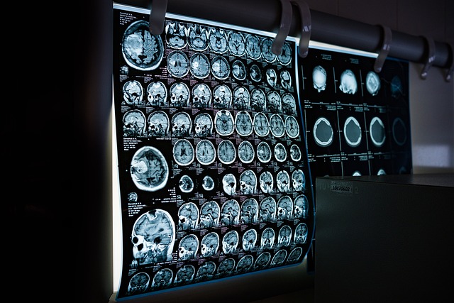
Alzheimer’s Diagnosis: Imaging Innovations
Diagnostic imaging has revolutionized modern medicine, enabling healthcare professionals to visualize internal body structures for accurate diagnoses and treatment planning. As technology advances, the field continues to evolve, promising improved patient care and more efficient healthcare delivery.

How is artificial intelligence transforming diagnostic imaging?
Artificial intelligence (AI) is rapidly changing the landscape of diagnostic imaging. Machine learning algorithms can now analyze medical images with incredible speed and accuracy, often outperforming human radiologists in certain tasks. AI-powered systems can detect subtle abnormalities that might be missed by the human eye, leading to earlier disease detection and more precise diagnoses.
These AI tools are particularly useful in analyzing large volumes of imaging data, such as in breast cancer screening programs. They can quickly flag potential areas of concern for radiologists to review, significantly reducing the time needed for image interpretation and potentially improving overall diagnostic accuracy.
What are the latest advancements in MRI technology?
Magnetic Resonance Imaging (MRI) continues to be a cornerstone of diagnostic imaging, and recent advancements have further enhanced its capabilities. High-field MRI scanners, operating at 7 Tesla and above, provide unprecedented image resolution and detail. This allows for more accurate visualization of small structures in the brain, joints, and other body parts.
Another significant development is the introduction of silent MRI technology. Traditional MRI scanners produce loud noises during operation, which can be distressing for patients. Silent MRI techniques dramatically reduce noise levels, improving patient comfort and potentially leading to better image quality by reducing motion artifacts caused by patient stress or discomfort.
How are imaging techniques improving Alzheimer’s detection?
Advanced imaging techniques are playing a crucial role in the early detection and diagnosis of Alzheimer’s disease. Positron Emission Tomography (PET) scans using specialized tracers can now visualize the accumulation of amyloid plaques and tau tangles in the brain – hallmarks of Alzheimer’s disease – years before symptoms appear.
Functional MRI (fMRI) is another powerful tool in Alzheimer’s research and diagnosis. By measuring blood flow changes in the brain, fMRI can reveal alterations in brain activity and connectivity associated with early-stage Alzheimer’s, potentially allowing for earlier intervention and treatment.
What new radiology solutions are enhancing diagnostic accuracy?
Dual-energy CT (DECT) is an innovative technology that’s gaining traction in radiology departments worldwide. By using two different energy levels, DECT can provide more detailed information about tissue composition and function compared to conventional CT scans. This technique is particularly useful in characterizing kidney stones, detecting bone marrow edema, and differentiating between benign and malignant tumors.
Another exciting development is the integration of 3D printing with diagnostic imaging. Radiologists can now create accurate 3D models of patients’ anatomy based on CT or MRI scans. These models are invaluable for surgical planning, patient education, and even as guides during complex procedures.
How is diagnostic imaging becoming more patient-friendly?
The push towards patient-centered care has led to significant improvements in the diagnostic imaging experience. Ultra-fast MRI protocols have been developed that can complete full-body scans in as little as 15 minutes, reducing patient discomfort and improving throughput in busy radiology departments.
Portable imaging devices are also becoming more common, allowing for bedside imaging in intensive care units or emergency departments. These mobile units reduce the need to transport critically ill patients, improving safety and efficiency.
What role does AI play in streamlining radiology workflows?
AI is not only enhancing image analysis but also optimizing radiology workflows. Intelligent scheduling systems can prioritize urgent cases and allocate resources more efficiently. Natural language processing algorithms can assist in report generation, allowing radiologists to dictate findings which are then automatically transcribed and formatted.
AI-powered quality control systems can also flag potential errors or inconsistencies in imaging studies before they are finalized, reducing the risk of misdiagnosis and improving overall patient care.
The field of diagnostic imaging is rapidly evolving, driven by advances in AI, MRI technology, and novel radiology solutions. These innovations are enhancing diagnostic accuracy, improving patient experiences, and streamlining clinical workflows. As research continues, we can expect even more groundbreaking developments that will further transform healthcare delivery and improve patient outcomes.
This article is for informational purposes only and should not be considered medical advice. Please consult a qualified healthcare professional for personalized guidance and treatment.
The shared information of this article is up-to-date as of the publishing date. For more up-to-date information, please conduct your own research.


